Vascular Cell Biomechanics and Mechanotransduction
Cardiovascular disease is the world’s leading cause of death, accounting for approximately one-third of all deaths. Among the most serious of these diseases is atherosclerosis, the buildup of plaque in the arterial walls. Although many of the risk factors for this disease affect the whole body, atherosclerosis is most likely to develop in specific local areas of the vasculature.
To better understand the development of cardiovascular disease, we use in vitro testing to apply mechanical forces to endothelial cells to replicate the mechanical environments present in healthy or diseased arteries. By studying the cellular response, we can evaluate the role of mechanical forces such as stretching and fluid shear stress in disease development.
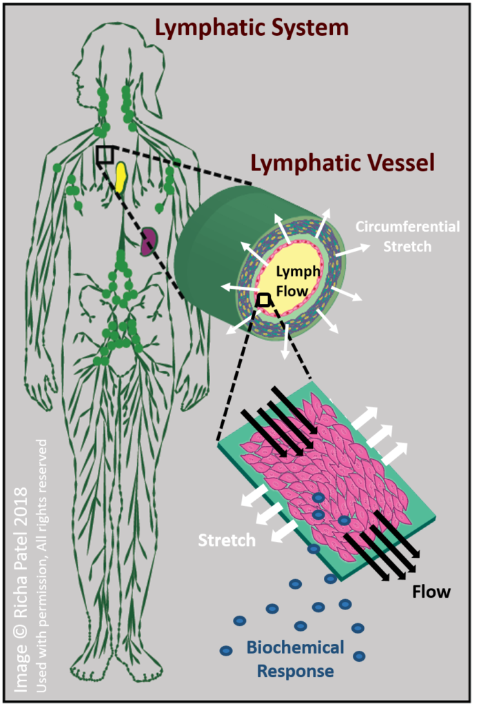
Lymphatic Cell Biomechanics and Mechanotransduction
The lymphatic vessels perform vital transport roles, such as removing excess fluid from the tissues and pumping it back into the circulatory system. Inadequate lymphatic fluid transport leads to edema (swelling), a painful and difficult to treat condition affecting millions worldwide. To maintain fluid transport, many lymphatic vessels are lined with smooth muscle which contracts rhythmically. The strength and frequency of contraction varies widely based on mechanical conditions in the vessel, and the mechanisms regulating this behavior are not well understood.
To better understand the self-regulating mechanisms of lymphatic transport, we use in vitro testing to apply mechanical forces to lymphatic endothelial cells to replicate the unique mechanical environments found in the lymphatic vessels. Studying the cellular response will help us evaluate the role of mechanical forces such as stretching and fluid shear stress in mechanoregulation of lymphatic transport.
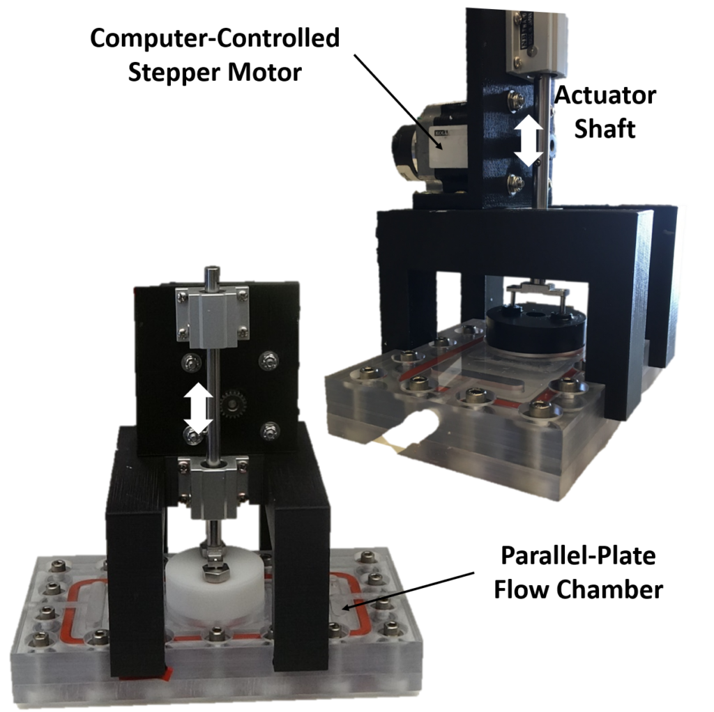
In Vitro Bioreactor Testing
Our lab has developed and built multiple custom bioreactors for culturing cells under controlled levels of fluid shear stress and/or stretching. This allows us to replicate many different scenarios of interest, such as the unique mechanical environment of the lymphatic vessels, or the altered direction of shear stress with respect to stretching similar to what is found in arterial areas prone to atherosclerosis.